Saturday, March 31, 2012

Gamma knife Radiosurgery for Glomus Jugulare Tumor.

This 24 years old lady presented with h/o vertigo since 10 years, bilateral tinnitus more on right side since 2 years, headache since one year. She had loss of right gag and palatal reflexes, atrophied tongue on right side with fasciculation and right deviation.
.Patient was treated with marginal dose of 14 Gy with 43% isodose line was prescribed for the target volume 20.3 cc; multiple iso centers with 18, 14 and 8 mm collimators were used in APS and trunnion mode.
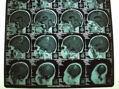
Pre Gamma Knife radiosurgery.
Glomus jugulare Tumor.
Volume 20.3 cc.

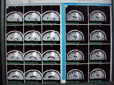
Follow up CT images Obtained  18 months after GKRS, showing tumor volume 5.2 cm3.
There is remarkable clinical improvement especially in tinnitus and hoarseness.

Tuesday, February 28, 2012

Gamma Knife Radiosurgery in Low Grade Glioma, decrease in T2 High Signal Volume


MRI brain with contrast revealed  non enhancing mass in the right parietal para falcine region over motor strip with high T2 signals.  MR Spectroscopy revealed neoplastic activity within the lesion. He had Gamma Knife Radiosurgery with following prescription.
Target
                Location
           Prescription
Volume
A
Right parietal Glioma
       12 Gy @ 50%
 17.4 cm³


Low Grade Glioma: Follow up at 3 months shows about 25 % decrease in T2 high signal volume.
Pt. is clinically in static condition with fits controlled on medicine.


Wednesday, January 18, 2012

Update on Oligodendroglioma GIII,Initially posted last year.


Oligodendroglioma, Grade III (Recurrent)Before Gamma Knife


This 45 years old Lady had undergone left frontal Craniotomy in November 2009 on diagnosis of left frontoparietal  tumor,. On histopathology it was Anaplastic Oligodendroglioma, WHO grade III. She had tonic clonic fits and right hemiparesis. she had received Radiotherapy to left hemisphere using left lateral and posterior fields on 6Mv X-ray beam.  Total dose of 60 Gy in Multiple fractions of 200 cGy had received until March 1, 2010. Temozolamide as conjoined chemotherapy used for 6 weeks during radiotherapy and 3 cycles after one month of radiotherapy.  

On referral, for She had presented progressive recurrence of right hemiparesis and fits since first week of April 2010. On Clinical examination, she had House Brackmann right facial palsy grade I-II and right hemiplegia. MRI brain with contrast had revealed multiple heterogeneous rim enhancing mass in the left parietal region with significant perilesional edema.  Patient referred us for management with GKSRS. Risk of GKSRS explained. These agreed upon wished to proceed. She had treatment with following prescription.
Target
                             Location
                   Prescription
       Volume
A
Left parietal Glioma
  12.0 Gy @ 50%
33.6 cm³
B
Left parietal Glioma lateral part
  12.0 Gy @ 40%
 3.5 cm³

Multiple isocenters with 18 & 8 mm collimator used in APS mode. She discharged on tapering doses of Dexamethasone and advised follow up after 3 months.

Follow up at 4 months significant  shrinkage of the tumor mass.


Follow up at 9 months shows further decrease in enhancment.

one and half year follow up:
Same clinical and radiological status as seen in last follow up.

Wednesday, January 11, 2012

Recurrent Meningeal Hemangiopericytoma, 50% resolution at 8 month follow up.


This 43 years old  gentleman from Gujranwala went through excision of posterior fossa tumor in 2008 along with a VP shunt . He presented with complain of headache, vertigo, and imbalance.

 On referral for Gamma Knife, MRI brain with contrast shows the recurrent multilobulated midline extra axial mass  occupying the supracerebellar cistern, encroaching into quadrigeminal cistern besides bulging into upper 4th ventricle.Mass was compressing upon adjacent faces of occipital lobes and superior vermis / adjacent cerebellum. He was treated with 14 Gy at 50% IDL for a Vol. of 27.8 cc. with multiple isocenters.


At GKRS


 Follow up at 8 months shows about 50% resolution. All his symptoms are improved.


After 8 months.

Meningeal Hemangiopericytoma is a rare and aggressive CNS tumor that exhibits a high incidence of local recurrence and distant metastasis.It arises from Zimmerman pericytes in the capillary wall.Till 2001 only 100 caes were reported.


Friday, December 2, 2011

Ependymoma, Two years follow up.

17 yrs, Female.
with persistent headache.
Known case of ITP.
MRI showed
 non enhancing mass 
periventricular area 
more on Lt. side.
Burhole biopsy 
showed
Ependymoma Gr.II.

Complete resolution after one year of Gamma knife Treatment.

                       At GKRS                             After one year                              After Two years


Monday, September 26, 2011

Cavernous sinus hemangioma, total resolution at 6 months.


Twenty five year old lady with restricted Rt.eye movements towards nasal side,
Drooping of rt. eyelid and mid dilated pupil.

MRI showed 2.7+2.9+3.4 cm homogenously enhancing solid mass just above
 rt. Cavernous sinus in right side of supra sellar cistern abutting anteromedial
Surface of rt. Temporal lobe, undersurface of thalamus and anterior portion of
right temporal lobe, undersurface of thalamus and anterior portion of rt. half of
midbrain, encasing more than 70% of circumference of top of basilar artery,
causing mild mass effect on pituitary stalk and optic chiasm on rt. Side.


6 month post GKRS
12 Gy at 50 % isodose line.
No Ptosis
Still has devation of rt. eyeball.

Thursday, August 18, 2011

Craniopharyngioma, complete resolution at one year follow up.


This 32 years old male is a diagnosed case of Craniopharyngioma with the diminished vision more on left side, headache and  a single episode of generalized fit since 2 and half years. Under went right frontal craniotomy on 2nd July 2007. MRI scan and CT scans showed residual/recurrent Craniopharyngioma. Patient was treated with 12 GY at 50% isodose line,  for the target volume 7.1 cc, multiple isocenters with 4 and 8mm collimators were used in APS mode.
Hostgator promotional code

F.up images after one year showing complete resolution of 
craniopharyngioma, clinically patient has normal vision and has no defecits.
< a href='http://www.ablogin.com/' > A Blogin Blog Directory< /a >