Showing posts with label Radiosurgery. Show all posts

Friday, March 29, 2024


 Gamma Knife Radiosurgery for Pituitary Adenoma.

Case Summary:

50 years male from Baluchistan, underwent trans sphenoidal excision of adenoma  in March 19, 2022 presented with complaints of progressive loss of right vision for 1 year & decrease left vision for 1 year. Patient has no history of chemotherapy and radiation therapy. On clinical examination pt. is blind from right eye with left temporal 
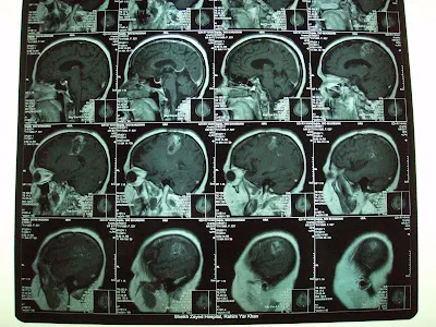
defect. MRI brain with contrast dated January 06, 2023 shows heterogeneous enhancing abnormal signal intensity mass lesion seen in sellar & supra sellar region suggestive of adenoma. Histopathology dated April 05, 2022 shows adenoma. Perimetry dated January 05, 2023 shows left temporal defect. Hormones profile within normal range. Risks of GKSRS have been explained. Written and valid consent obtained to proceed.


Follow up 
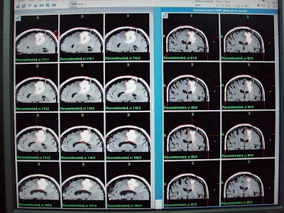
Comparative Study December 15, 2023:

Patient visited the gamma knife center for the first follow up presented with recent MRI brain with contrast dated December 14, 2023 shows further regression in the size of tumor from 4.6cc to 3.6 cc when compared with previous MRI brain with contrast dated July 04, 2023, consistent with good response to Gamma Knife Therapy


Thursday, June 28, 2012

Pineal region Tumor. Update at 2 years follow up.


This is a young man of 26 years  with c/o headache vomiting and blurred vision.

MR imaging showed an enhancing mass in the pineal region and obstructive hydrocephalus.
MR Specrtroscopy revealed alow NAA and high choline.
V.P shunt was placed and he was treated with Gamma Knife.It was a single day treatment as usual and patient was discharged next day.
At 6 months he is back to his normal life style and the Follow up MRI shows 90% resolution of the said tumor. Now at 2 years follow Pt. is fine with consistent resolution.
Resolution at 6 Month.
Further Resolution at one year F.up.

Further Resolution at one year F.up.
Consistent Resolution at 2 years follow up.


Tuesday, February 28, 2012

Gamma Knife Radiosurgery in Low Grade Glioma, decrease in T2 High Signal Volume


MRI brain with contrast revealed  non enhancing mass in the right parietal para falcine region over motor strip with high T2 signals.  MR Spectroscopy revealed neoplastic activity within the lesion. He had Gamma Knife Radiosurgery with following prescription.
Target
                Location
           Prescription
Volume
A
Right parietal Glioma
       12 Gy @ 50%
 17.4 cm³


Low Grade Glioma: Follow up at 3 months shows about 25 % decrease in T2 high signal volume.
Pt. is clinically in static condition with fits controlled on medicine.


Wednesday, January 18, 2012

Update on Oligodendroglioma GIII,Initially posted last year.


Oligodendroglioma, Grade III (Recurrent)Before Gamma Knife


This 45 years old Lady had undergone left frontal Craniotomy in November 2009 on diagnosis of left frontoparietal  tumor,. On histopathology it was Anaplastic Oligodendroglioma, WHO grade III. She had tonic clonic fits and right hemiparesis. she had received Radiotherapy to left hemisphere using left lateral and posterior fields on 6Mv X-ray beam.  Total dose of 60 Gy in Multiple fractions of 200 cGy had received until March 1, 2010. Temozolamide as conjoined chemotherapy used for 6 weeks during radiotherapy and 3 cycles after one month of radiotherapy.  

On referral, for She had presented progressive recurrence of right hemiparesis and fits since first week of April 2010. On Clinical examination, she had House Brackmann right facial palsy grade I-II and right hemiplegia. MRI brain with contrast had revealed multiple heterogeneous rim enhancing mass in the left parietal region with significant perilesional edema.  Patient referred us for management with GKSRS. Risk of GKSRS explained. These agreed upon wished to proceed. She had treatment with following prescription.
Target
                             Location
                   Prescription
       Volume
A
Left parietal Glioma
  12.0 Gy @ 50%
33.6 cm³
B
Left parietal Glioma lateral part
  12.0 Gy @ 40%
 3.5 cm³

Multiple isocenters with 18 & 8 mm collimator used in APS mode. She discharged on tapering doses of Dexamethasone and advised follow up after 3 months.

Follow up at 4 months significant  shrinkage of the tumor mass.


Follow up at 9 months shows further decrease in enhancment.

one and half year follow up:
Same clinical and radiological status as seen in last follow up.