Thursday, August 24, 2017
Wednesday, December 28, 2016
Gamma Knife Radiosurgery for Recurrent Medulloblastoma
This 7 years old boy from Faisalabad, biopsy proven Medulloblastoma WHO
G-IV, had undergone insertion of VP Shunt on October 01, 2013 & excision of
SOL on October 16, 2013 presented with complains of headache, ataxia &
double vision since 2013 which improved after surgery. Patient had whole brain radiotherapy, in 30 fraction, from November 2013 to December
2013. Seven cycles of Chemotherapy completed in September 2014. On clinical examination
patient has no obvious neurological deficit. MRI brain with contrast dated
January 08, 2015 shows interval increase in dural based nodular lesion rising from the left tentorial leaf, suggestive of
recurrence of disease. Patient referred us for Gamma Knife. Patient has following
treatment,
Target
|
Location
|
Prescription
|
Volume
|
A
|
Recurrent
Medulloblastoma
|
16.0 Gy @ 50 %
|
5.0 cm³
|
Multiple
isocenter with 18 & 08 mm collimator used in APS mode. The dose was
consulted with the Radiation Oncologist and was delivered with Leksell Gamma
Unit 4-C.
At Gamma Knife Treatment.
Friday, December 18, 2015
Saturday, November 7, 2015
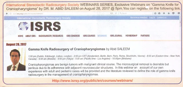
Talk on Managment of Craniopharyngiomas.
National Neurology Conference.2014
https://www.facebook.com/nmihospital/videos/914821428603374/
Wednesday, December 31, 2014
Gamma knife Radiosurgery for Residual Pituitary Adenoma.
48 years old gentleman with history of progressive visual loss ( finger counting only) and chronic headaches.
Status post Transphenoidal Adenectomy.
4.8 c.cm treated with 17 Gy @ 46% isodose line..May 15 2010.
Follow up images in Oct. 2014 show complete resolution of pituitary adenoma with no visual deficits.
Friday, April 19, 2013
Role of Gamma Knife Radiosurgery in Multimodality Management of Craniopharyngioma.
Article
Role of Gamma Knife Radiosurgery in Multimodality Management of Craniopharyngioma.
Department of Neurosurgery, Pakistan Gamma Knife and Stereotactic Radiosurgery Center, NeuroSpinal and Medical Institute, 100/1 Mansfield Street, M.A. Jinnah Road, Sadder, Karachi, 74400, Pakistan, .
Acta neurochirurgica. Supplement 01/2013; 116:55-60. DOI:10.1007/978-3-7091-1376-9_9
Source: PubMed
ABSTRACT Objective: This retrospective study evaluated the efficacy and safety of the use of Gamma Knife Radiosurgery (GKS) along with other surgical procedures in the management of craniopharyngioma. Methods: Thirty-five patients (17 children and 18 adults) with craniopharyngioma were treated with GKS between May 2008 and August 2011. The age of the patients ranged from 2 to 53 years (mean 20 years). There were 26 males and 9 females. Craniopharyngiomas were solid in 7 patients, cystic in 4, and mixed in 24. Tumor size ranged from 1 to 33.3 cm(3) (mean 12 cm(3)). The prescription dose ranged from 8 to 14 Gy (mean 11.5 Gy). Maximum dose ranged from 16 to 28 Gy (mean 23 Gy). Before GKS 11 patients underwent subtotal resection of the neoplasm, 2 - neuroendocopic fenestration of the large cystic component, and 10 - stereotactic aspiration of the neoplastic cyst content. Results: The length of follow-up period varied from 6 to 36 months (mean 22 months). The tumor response rate and control rate were 77.1 % and 88.5 %, respectively. Clinical outcome was considered excellent in 10 cases, good in 17, fair in 4, and poor in 4. No one patient with normal pituitary function before GKS developed hypopituitarism thereafter. Deterioration of the visual function after treatment was noted in one patient. Conclusion: After GKS tumor control can be achieved in significant proportion of patients with craniopharyngioma. Treatment-related neurological morbidity in such cases is rare. Therefore, radiosurgery may be considered useful for management of these tumors.
Subscribe to:
Comments (Atom)


