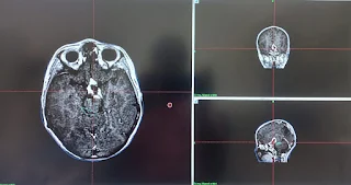
This 8 years child underwent excision of supra sellar SOL & insertion of VP shunt in April 24, 2022 presented with complaints of headache with vomiting before surgery & decrease left vision for 1 month. CT scan brain plain dated April 19, 2022 shows a large hyper dense lesion seen in supra sellar region with multiple peripheral calcific foci causing compression effect over optic chiasm with hydrocephalus suggestive of craniopharyngioma.
Follow-up Comparative Study after 6months :
Patient visited the gamma knife center for first routine follow up presented with improvement of left vision.
Follow up MRI brain with contrast dated December 29, 2022 shows significant regression in tumor volume from 41 cc to 5.7 cc.
Follow-up Comparative Study after 1 year:
Patient visited the gamma knife center for first routine Follow up presented with good health and no symptoms.Follow up MRI brain with contrast dated July 06, 2023 shows significant regression in tumor volume as reported previously.


.jpeg)